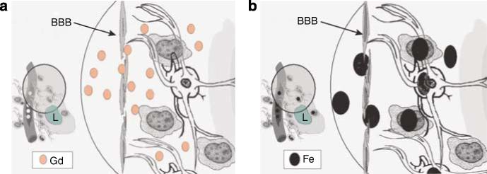
Figure 2. Schematic distribution comparing Gd versus USPIO crossing the BBB to tumor.

(a) Shows Gd chelates (abbreviated Gd in the figure) crossing from the intravascular compartment through a defective BBB (arrow) to the interstitial space. GBCA, which vary in propensity to release Gd, and thereby possibly risk of NSF (see text), remains in the extracellular space at the time of imaging (minutes after infusion). Macrophages, reactive astrocytes are shown around the lesion (L) but GBCA does not enter these cells. During its short half-life (less than 1 h), Gd rapidly crosses the BBB to produce a hyperintense signal on T1-weighted sequence on MR. In CKD, Gd-chelated contrast is not cleared rapidly and can be demonstrated in tissues.22 (b) Ferumoxtran-10 (Fe in black) and other ultra small iron oxides are much larger particles compared to GBCAs; these particles will slowly cross the defective BBB (arrow). Due to its prolonged half-life of 25−30 h particles of ferumoxtran-10 will progressively accumulate in the interstitial space where they will be trapped by reactive astrocytes and macrophages for days (up to 7 days). The intracellular metabolism of ferumoxtran-10 will take place in the lysosome of these cells where free iron will be released to the extracelullar space. Reactive astrocytes, macrophages, and other inflammatory cell are seen in different diseases affecting the CNS, among these stroke, neoplasm, and demyelinating processes. Figure modified from Murillo et al.13 (reproduced with permission of Expert Reviews Ltd).
