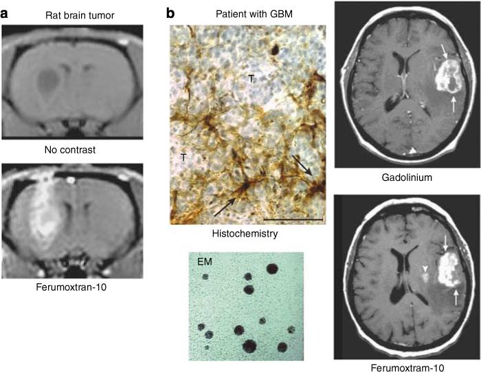
Figure 3. Nanoimaging: from bench to bedside.

(a) Rat brain tumor before and after USPIO MR contrast. (b) Post-GBCA T1-weighted MRI shows a large left frontoparietal-enhancing tumor (arrows). At 24 h post–ferumoxtran, T1-weighted MRI shows intense enhancement in the left frontoparietal tumor (arrows) and a new lesion medial to the main tumor mass in the putamen (arrowhead). Cellular iron staining at the tumor–reactive brain interface shows iron uptake by the parenchymal cells with fibrillar processes (arrows) rather than by the round tumor cells (T). EM shows electron dense core of USPIO.1 (Reproduced from AJNR Am J Neuroradiol 2002; 23:510−519 with permission of Lippincott, Williams & Wilkins, 2008.)
