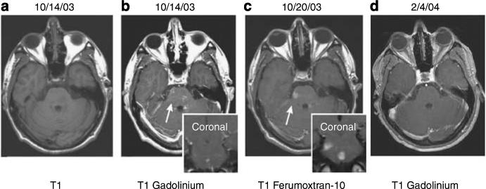
Figure 4. Patient with acute disseminated encephalomyelitis (ADEM).

Axial T1-weighted MRI images without (a) and with (b) GBCA show faint, subtle enhancement in multiple brainstem lesions. At 6 days later (c) significant, more prominent, larger ferumoxtran-10 enhancement can be seen on the same site. At 3 months later (d), the lesions no longer enhance on T1-weighted images with GBCA.2 (Reproduced from AJNR Am J Neuroradiol 2005; 26:2290−2300 with permission of Lippincott, Williams & Wilkins, 2008).
