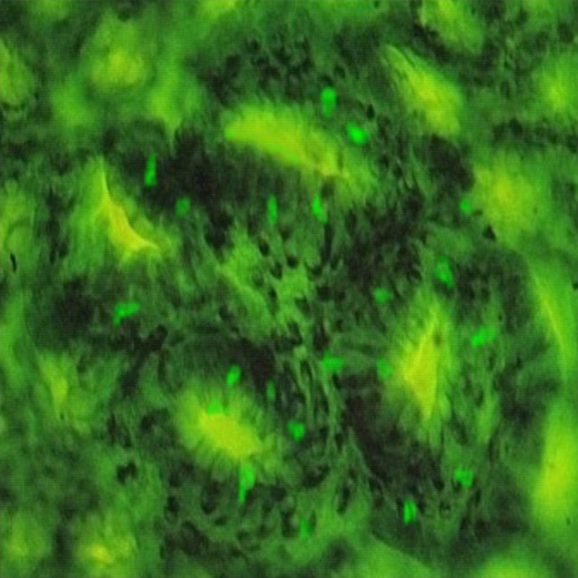
Figure 3. Fluorescein dextran labeled cells on top of a four-crypt ACF.

Fluorescence micrograph of colon mucosa (magnification × 400). The rat was sacrificed 130 d after an AOM injection, and 30 h after a 250 mg fluorescein dextran gavage (this rat did not receive PEG).
