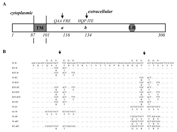
Fig. 1.
A. Schematic diagram showing ST3 cleavage sites between the transmembrane domain (TM) and the laminin binding sequence (LB) of LR (27). ST3 cleaves LR between A115 and F116 (site a), and P133 and I134 (site b) and these two cleavage sites by ST3 are indicated by two arrows (Based on (27)).
B. LR mutants used in this study.

