
Figure 4.
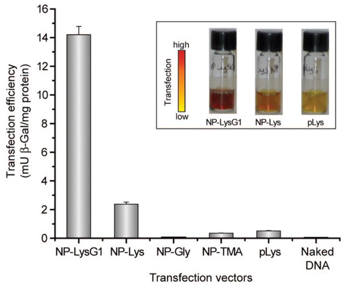
Enhanced transfection using NP–LysG1 and NP–Lys relative to positive controls, NP–TMA, and polylysine (pLys). No appreciable enzyme activity was observed in the absence of vectors. Inset shows solution colors during β-Gal activity assay performed after transfection. The color changes from yellow (substrate) to red (product) in the presence of active enzyme.