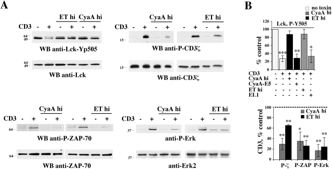
Figure 5. Immunosuppressive concentrations of CyaA or ET prevent initiation of TCR signaling.
(A) Top left, Immunoblot analysis, using a phosphospecific antibody, of Lck phosphorylation on the inhibitory C-terminal tyrosine residue (Y505) in postnuclear supernatants from PBL activated for 1 min by CD3 cross-linking in the presence or absence of either 45 nM CyaA or 110 nM ET (CyaA hi, ET hi). Top right, Immunoblot analysis, using an anti-phosphotyrosine antibody, of CD3ζ specific immunoprecipitates from PBL treated as above. Bottom, Immunoblot analysis, using phosphospecific antibodies, of ZAP-70 (left) and Erk1/2 (right) phosphorylation in postnuclear supernatants from PBL activated for 1 min (ZAP-70) or 5 min (Erk1/2) by CD3 cross-linking in the presence or absence of 45 nM CyaA or 110 nM ET (CyaA hi, ET hi). (B) Quantification by laser densitometry of the relative levels of Lck (phosphorylation in unstimulated cells taken as 100%), or CD3ζ, ZAP-70 and Erk1/2 phosphorylation (phosphorylation in anti-CD3 stimulated cells taken as 100%, indicated as a dotted line) in PBL activated by CD3 cross-linking in the presence of 45 nM CyaA or 110 nM ET (CyaA hi, ET hi) (n≥3). Where indicated, cells were activated in the presence of the adenylase cyclase deficient CyaA and ET mutants (45 nM CyaA-E5, 110 nM EL1) for 2 h or 6 h, respectively (n = 2). ***P≤0.001; **P≤0.01; *P≤0.05. Error bars, SD.

