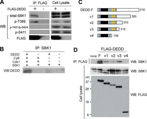
FIGURE 2.
DEDD forms a complex with S6K1 and Cdk1 via its proline-rich region. A, co-immunoprecipitation (IP) of FLAG-tagged DEDD (FLAG-DEDD) and S6K1, using anti-FLAG antibody. Precipitates were analyzed for the indicated items. Western blots (WB) of cell lysates for respective subjects are also presented. Note in 293T cell lysates, protein blot for total S6K1 using a mouse monoclonal antibody (clone 16; BD Biosciences) reveals double bands, as previously described (40, 41). B, in vitro binding assay using recombinant proteins. C, schematic diagram of the structure of DEDD variants. Light blue region, nuclear localizing signal; black region, DED domain; orange region, proline-rich region. The numbers indicate amino acids starting from the first methionine. D, co-immunoprecipitation of S6K1 and FLAG-tagged DEDD variants, using an anti-FLAG antibody.

