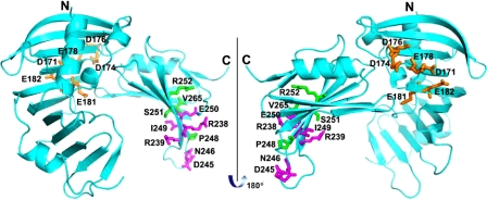
FIGURE 9.
Crystal structure of RseB with alanine and cysteine substitutions marked. The three groups of substitutions shown in color are depicted using a ball and stick representation: 1) C-terminal residues R238, R239, D245, N246, I249, and E250, which were mutated to alanine are colored magenta, 2) N-terminal residues D171, D174, D176, E178, E181, and Q182, which were mutated to alanine are colored orange, and 3) C-terminal residues P248, S251, R252, and V265, which were mutated to cysteine are colored green.

