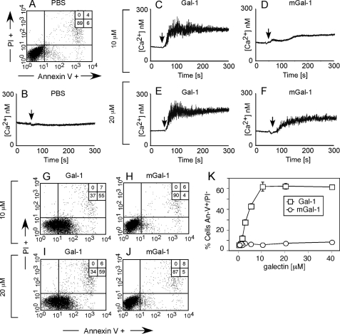
FIGURE 1.
mGal-1 induces proximal signaling events in leukocytes but fails to induce PS exposure. A, HL60 cells treated with PBS for 4 h were stained with annexin-V fluorescein isothiocyanate and propidium iodide (PI) to detect PS exposure followed by flow cytometric analysis. B, cells were loaded with Fluo-4 and analyzed for changes in intracellular Ca2+ using a fluorometer following addition of PBS at the indicated time (vertical arrow). C–F, HL60 cells were loaded with Fluo-4 as in B, followed by addition of: 10 μm Gal-1 (C), 10 μm mGal-1 (D), 20 μm Gal-1 (E), or 20 μm mGal-1 (F) as indicated by the arrows. G-J, HL60 cells treated with 10 μm Gal-1 (G), 10 μm mGal-1 (H), 20 μm Gal-1 (I), or 20 μm mGal-1 (J) for 4 h were used to label cells with annexin-V fluorescein isothiocyanate and PI to detect PS exposure followed by flow cytometric analysis. K, HL60 cells were treated with the indicated concentrations of Gal-1 or mGal-1 for 4 h followed by detection for PS exposure.

