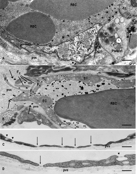
FIGURE 1.
Morphological alterations induced by PAF in mouse lung microvessels. A, opening of large gaps (∼1 μm) in the endothelium of a lung venule. Platelet and Monastral Blue particles (arrows) have escaped through the interendothelial gap and are retained in the perivascular space (pvs) by the basement membrane. Note the presence of red blood cells (RBC) and Monastral Blue tracer within the vascular lumina. Bar, 350 nm. B, interendothelial gaps encountered in the lung capillaries. A gap of ∼2 μm was crammed with fingerlike projections (F1 and F2) in the endothelium of a capillary. Note the escaped particles of Monastral blue (arrows) and the presence of long cellular extensions belonging to one cell (F1) as well as the existence of sectioned intercellular projections (F2) that could belong to any of the two ECs facing the gap. In both A and B, the ECs facing the open gaps have a higher than normal number of intracellular vacuoles (v). Bar, 250 nm. C and D, typical fenestrae (arrows) found in venular (C) or capillary (D) endothelium. After PAF administration, classic fenestrations closed by diaphragms were found both in the capillary and venular end of the murine lung vascular bed. There is a tendency for PAF-induced fenestrae to be grouped in the venular endothelium (C). Also note that some of the luminally opened vesicles (arrow-heads) in C and D are provided with a diaphragm. Bar, 175 nm; the micrographs are representative of five experiments.

