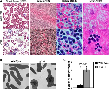
FIGURE 4.
A, histology of blood, spleen, and liver, from heterozygous human γβ0 globin KI mice shows β thalassemic phenotypes. Wright-Giemsa stained peripheral blood smears of wild-type control and heterozygous γβ0 globin KI mice. Typical red blood cell targeting, hypochromia, and anisopoikilocytosis are observed in thalassemic human γβ0 globin KI mice. The normal red (erythroid) and white (lymphoid) pulp sections seen in the wild-type spleen were replaced by an expanded erythroid compartment in the γβ0 globin KI mice compared with the wild-type control. Livers of thalassemic γβ0 globin KI mice contain clusters of erythroid cells indicative of extramedullary hematopoiesis. Image magnifications are shown in parentheses. Slides were analyzed on Nikon Eclipse E800 or TE2000 microscopes (Nikon, Tokyo, Japan). Image magnifications are shown in parentheses. B, electron microscopy of circulating red blood cells from wild type and γβ0 globin KI adult mice demonstrating α globin chain inclusions (scale bar, 1 μm). C, comparison of spleen size as a percentage of body weight of wild type and γβ0 globin KI mice show a significant splenomegaly in the thalassemic mice (n ≥ 8 in each group).

