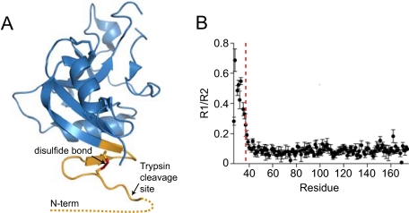
FIGURE 5.
The HIP/PAP N terminus is flexible. A, ribbon diagram of the pro-HIP/PAP crystal structure (RCSB accession: 1UV0) (15). The disordered N-terminal 10 amino acids are indicated by a dotted line, and the locations of the N-terminal disulfide bond (red) and the trypsin cleavage site are indicated. B, experimental 15NR1 and R2 relaxation rates were determined for pro-HIP/PAP, and a plot of the R1/R2 ratio is shown. The location of the Arg37–Ile38 trypsin site is indicated by a dashed red line.

