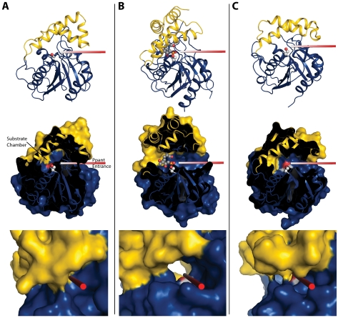
FIGURE 4.
Cartoon and surface diagrams in equivalent orientations for RifR TEII (A), Pik TEI (Protein Data Bank code 2HFJ) with affinity label (B), and SrfTEI (Protein Data Bank code 1JMK) (C). The rods show the Ppant entrance path to the catalytic serine (shown in spheres). The middle panels show a cut-away surface diagram in the same orientation as the top panels. Access to the active site for RifR TEII is blocked by the lid helices (yellow). The bottom panels are close-up views along the Ppant entrances, showing the closed entrance in RifR, the tunnel-like entrance characteristic of dimeric PKS TEIs (Pik TEI) and trough-like entrance of the monomeric NRPS TEI (SrfTEI in the open form).

