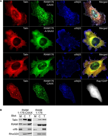
FIGURE 4.
Expression of RIAM-(1–176)-CAAX targets talin to integrins in the plasma membrane. A, A5 cells transiently expressing mCherry-talin and GFP-tagged RIAM-(1–176)-CAAX, RIAM-(1–176)-A-SAAX, or RIAM-(1–176) (top three panels), or with RIAM-(1–176)-CAAX and HA-Rap1GAP (bottom panel) were adhered to fibrinogen-coated coverslips and stained to visualize integrin αIIbβ3 and Rap1GAP. Depicted are the localization of αIIbβ3(blue), talin (red), RIAM (green), or Rap1GAP (white). Images shown are maximal projections of deconvoluted 0.1-μm z-section images of the entire cell volume. B, A5 cells expressing HA-talin and GFP-tagged RIAM-(1–176)-CAAX or RIAM-(1–176) were Dounce-homogenized, fractionated, and analyzed by Western blotting for the distribution of HA-talin and GFP-RIAM in membrane (M), cytosol (C), and total (T) fractions with the corresponding anti-HA or anti-GFP antibodies. In addition, integrin αIIb and RhoGDI were immunoblotted to validate separation of the membrane and cytosol fractions, respectively.

