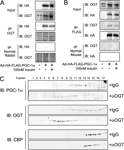
FIGURE 3.
PGC-1α interacts with OGT. A, Fao cells were infected with Ad-FLAG-PGC-1α and treated with insulin for 1 h prior to harvesting and OGT co-immunoprecipitation (IP). Immunoprecipitates were subjected to SDS-PAGE and immunoblotted (IB) for the presence of PGC-1α. Membranes were then stripped and blotted for OGT. Immunoblots are representative of three experiments. B, Fao cells were infected with Ad-FLAG-PGC-1α and treated with insulin for 1 h prior to harvesting and FLAG co-immunoprecipitation. Immunoprecipitates were subjected to SDS-PAGE and immunoblotted for the presence of OGT. Membranes were then stripped and blotted for PGC-1α. C, gel filtration chromatography (SMART system, Superdex 200 column, phosphate-buffered saline, 1% Nonidet P-40 buffer) of lysates from rat liver incubated on ice for 30 min with either normal IgG or anti-OGT antibodies (AL28). Fractions were subjected to SDS-PAGE and blotted using anti-PGC-1α, anti-OGT (DM-17), and anti CPB. Data are representative of two experiments.

