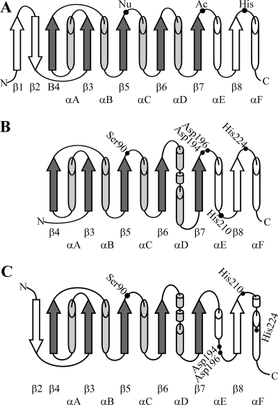
FIG. 2.
Topology of α/β hydrolases. Cylinders, α helices; arrows, β strands. Shading indicates the minimal set of α and β structures. (A) Canonical α/β hydrolase fold. Catalytic triad amino acids are indicated by dots. (B) Predicted topology of secondary structures of ScoT thioesterase representing model 1. (C) Predicted topology of secondary structures of ScoT tioesterase representing model 2. In panels B and C amino acids selected for site-directed mutagenesis are indicated by dots.

