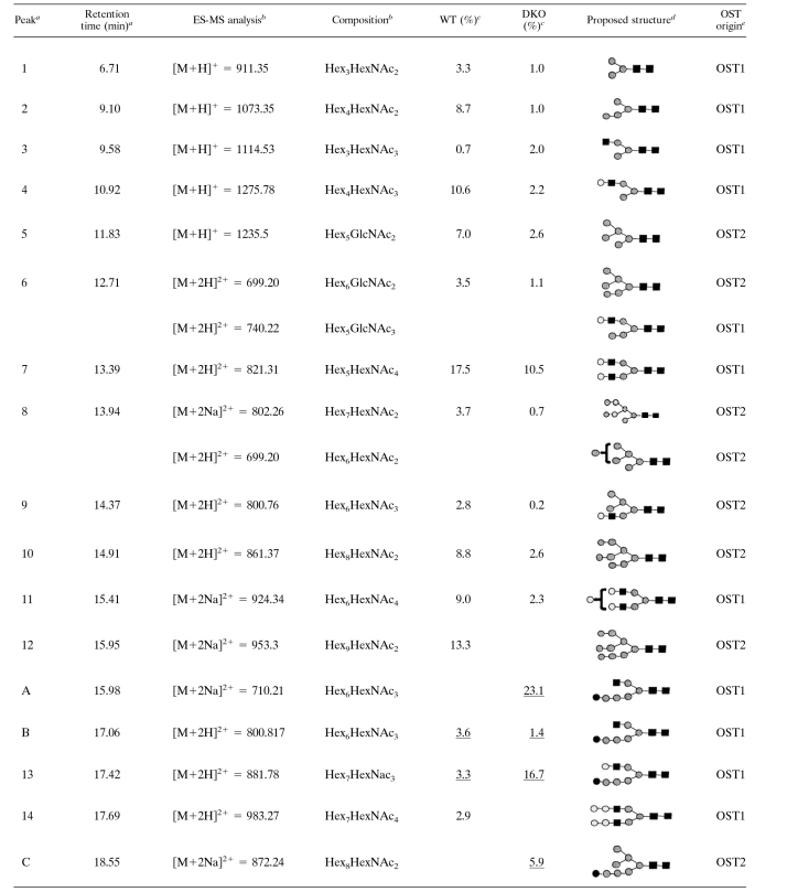
TABLE 1.
N-glycans expressed by wild-type and GII null mutant trypanosomes
The peak identifiers and retention times refer to the Dionex HPAEC chromatograms shown in Fig. 1C and D.
The positive-ion ES-MS analyses of the individual peaks revealed the main molecular species as being [M+H]+, [M+2H]2+, or [M+2Na]2+ ions from which the glycan compositions were deduced.
The proportions of each glycan are estimated from the areas of the peaks in Fig. 1C and D. WT, wild type. Underlined values indicate N-glycans retaining one terminal κ-Glc residue.
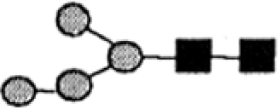
The proposed structures are based on the known compositions, the ES-MS-MS spectra of the principal ions shown in Fig. 1A and B, and literature precedent for T. brucei N-glycans with those compositions.
Likely origin of the N-glycan with respect to the OST (OST1 or OST2) (39) that will have transferred its Man5GlcNAc2 (OST1) or Man9GlcNAc2 (OST2) precursor.


