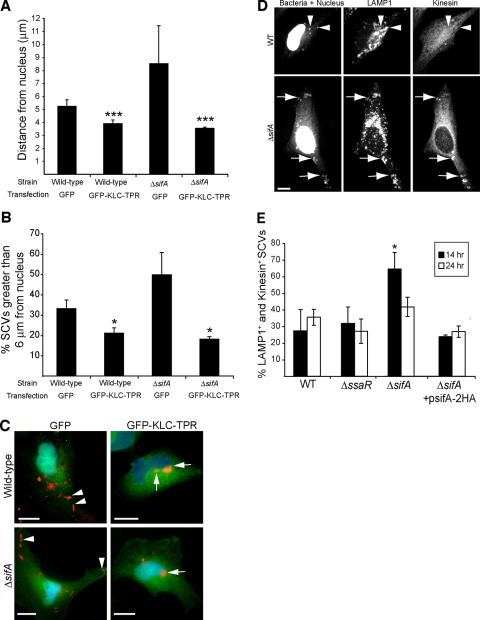
FIG. 3.
Kinesin is required for the centrifugal displacement of intracellular serovar Typhimurium at late time points postinfection. (A) HeLa cells were infected with WT or ΔsifA serovar Typhimurium, and subsequently transfected (2 h postinfection) with a plasmid encoding GFP alone or GFP-KLC-TPR that has dominant-negative activity against kinesin. Cells were fixed at 24 hpi and immunostained for serovar Typhimurium. The distances of bacteria to the nearest edge of the host cell nucleus were measured in transfected cells. The averages ± standard deviations for three separate experiments are shown. A triple asterisk indicates a significant difference from the respective GFP transfection control (P < 0.0.001), as determined by Kruskal-Wallis one-way ANOVA with Dunn's multiple comparison posttest. (B) Percentage of SCVs examined in panel A that were located >6 μm from their host cell nucleus. Averages ± standard deviations for three separate experiments are shown. The asterisk indicates a significant difference from the respective GFP transfection control (P < 0.05), as determined by a two-tailed, unpaired t test. (C) Positioning of WT and ΔsifA serovar Typhimurium bacteria (red) 24 hpi in HeLa cells transfected with GFP or GFP-KLC-TPR. Arrowheads indicate bacteria positioned toward the host cell periphery in GFP-transfected cells. Arrows indicate bacteria positioned next to the host cell nucleus in GFP-KLC-TPR-transfected cells. Size bar, 10 μm. (D) Typical kinesin labeling of WT (top panel) and ΔsifA (bottom panels) LAMP1+ SCVs in infected HeLa cells at 14 hpi. Arrows indicate distinct recruitment of kinesin to LAMP1+ SCVs of the ΔsifA strain. Arrowheads indicate colocalization of LAMP1 with WT bacteria, which do not display significant kinesin recruitment regardless of their position in the host cell. Cells were also DAPI stained. Size bar, 10 μm. (E) HeLa cells were infected with WT, ΔsifA, ΔssaR, or ΔsifA (psifA-2HA) strains of serovar Typhimurium for the times indicated. Cells were fixed and immunostained for LAMP1, bacteria, and kinesin. The percentage of LAMP1+ SCVs colocalizing with kinesin was determined for each strain. Values shown are the averages ± standard deviations of three separate experiments. The presence of kinesin on 100 LAMP1+ SCVs was determined for each strain at each time point in a given experiment. The asterisk indicates a significant difference between the indicated mutant strain and the corresponding WT control (P < 0.05), as determined by a two-tailed Mann-Whitney test.

