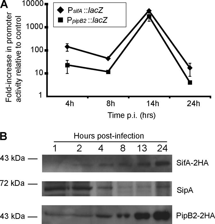
FIG. 5.

Analysis of promoter activity and protein expression levels of SifA, PipB2, and SipA. (A) HeLa cells were infected with serovar Typhimurium carrying chromosomal transcriptional lacZ fusions to either the sifA or pipB2 promoters (PsifA::lacZ or PpipB2::lacZ) or with the WT parental strain. At selected times, cell lysates were collected, and β-galactosidase activity was determined using a chemiluminescence assay. Light signal data were normalized to the numbers of CFU, based on dilution plating of lysates. Shown are the relative increases in β-galactosidase activity resulting from PsifA::lacZ or PpipB2::lacZ activity compared to the WT control infection. Values are the averages from two independent experiments and average deviations. (B) Immunoblot analysis of effector protein levels in infected HeLa cells. Cells were infected with ΔsifA (psifA-2HA) (top and middle blots) or ΔpipB2 (pPipB2-2HA) (bottom blot) strains of serovar Typhimurium. Whole-cell lysates were harvested at the indicated times and analyzed by immunoblotting samples that were loaded according to equal bacterial numbers. Top and middle blots show Western blotting to detect SifA-2HA and SipA, as indicated, from the same blot. At the bottom is a Western blot to detect PipB2-2HA.
