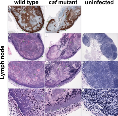
FIG. 4.
Differential histopathologies in the proximal lymph nodes of mice. Sections of infected (A to D, F to G, and I to J) and uninfected (E, H, and K) lymph nodes were stained by IHC using Y. pestis-specific antibody (A and B) or by H&E (C and K). Masses of bacteria, indicated by green arrowheads, strained brown by IHC and blue by H&E. Magnification, ×40 (A to E), ×100 (F to H), and ×400 (I to K).

