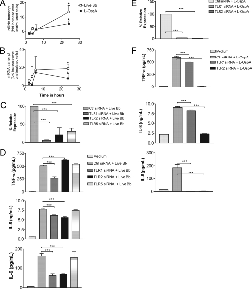
FIG. 3.
TLR1 or TLR2 siRNA transfection of THP-1 cells affects the production of inflammatory mediators in response to stimulation with live B. burgdorferi. Vitamin D3-treated THP-1 cells (3 × 10⁶ cells/ml) were incubated with supplemented antibiotic-free RPMI medium alone or with live B. burgdorferi at an MOI of 10 or with L-OspA at 1 μg/ml in complete medium. RNA samples were collected after 2, 4, 6, and 24 h of incubation and qRT-PCR was performed for TLR1 (A) and TLR2 (B) mRNA transcript levels. Results are presented as the increase over control (the level in unstimulated cells). Each data point represents the mean ± standard deviation (SD) of two independent experiments. (C) Cells were transfected and analyzed for TLR1, TLR2, and TLR5 knockdown as described in the legend to Fig. 2A. (D) Supernatants were analyzed by antibody capture ELISA for TNF-α, IL-8, and IL-6 production as described in the legend to Fig. 2C. (E) Cells were transfected with nontargeting control siRNA or with TLR1 or TLR2 siRNA and cultured in complete medium alone for 24 h. They were then stimulated with L-OspA (1 μg/ml) for an additional 24 h, and RNA was extracted from transfected cells and used in qRT-PCR to assess TLR1 and TLR2 knockdown as described in the legend to Fig. 2A. Results are the means ± SDs of three independent experiments. (F) Supernatants were analyzed by antibody capture ELISA for TNF-α, IL-8, and IL-6 production as described in the legend to Fig. 2C.

