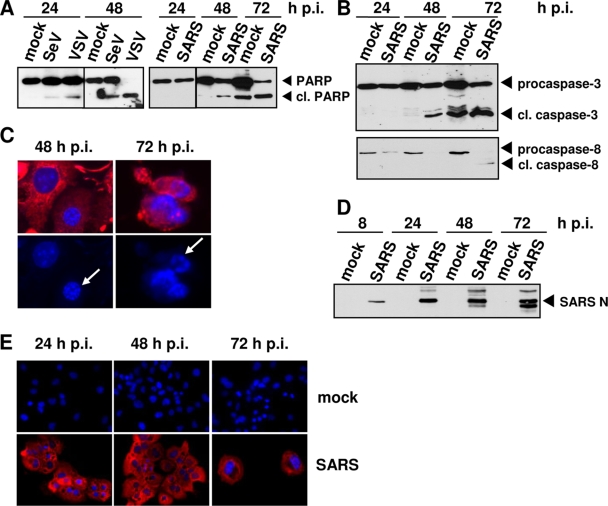
FIG. 1.
Induction of apoptosis in SARS-CoV-infected 293/ACE2 cells. (A) 293/ACE2 cells were infected with VSV (MOI, 0.01), SeV (20 hemagglutinating units), or SARS-CoV (MOI, 0.01). Protein extracts from samples collected at different time points p.i. were subject to Western blot analysis using anti-PARP antibody. (B) 293/ACE2 cells were infected with SARS-CoV as described for panel A, and the noncleaved and cleaved fragments of caspase-3 and caspase-8 were detected by Western blot analysis. (C) 293/ACE2 cells grown on coverslips were infected with SARS-CoV as described for panel A and subjected to immunofluorescence analysis using rabbit antiserum directed against the nucleoprotein of SARS-CoV (N) (red). DAPI (4′,6′-diamidino-2-phenylindole) staining (blue) visualizes cell nuclei. Chromatin condensation is indicated by white arrows. (D) 293/ACE2 cells were infected with SARS-CoV as described for panel A; lysed at 8, 24, 48, and 72 h p.i.; and subjected to Western blot analysis with anti-N antiserum. (E) Immunofluorescence analysis of noninfected and SARS-CoV-infected 293/ACE2 cells was performed as described for panel C.

