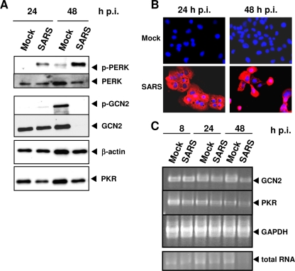
FIG. 6.
Induction of eIF2α-phosphorylating kinases during SARS-CoV infection. (A) 293/ACE2 cells were infected with SARS-CoV at an MOI of 0.01 and lysed at 24 h and 48 h p.i. Western blot analysis was performed with antibodies against β-actin and the phosphorylated (p) and nonphosphorylated forms of PERK, GCN2, and PKR. This experiment was repeated three times with similar outcomes. (B) 293/ACE2 cells grown on coverslips were infected with SARS-CoV as described for panel A. Immunofluorescence analysis using rabbit antiserum directed against the nucleoprotein of SARS-CoV (N) (red) revealed that 100% of the cells were infected. DAPI staining (blue) was performed to visualize cell nuclei. (C) Total RNA of 293/ACE2 cells infected with SARS-CoV as described for panel A was isolated at 8, 24, or 48 h p.i., and 5 μl of each RNA sample was subjected to RT-PCR analyses to determine GCN2-, PKR-, and GAPDH-specific mRNA levels (30 cycles). In addition, 1 μl of each total cellular RNA sample was analyzed by agarose gel electrophoresis. The 18S rRNA band is shown and indicates the amount of total cellular RNA.

