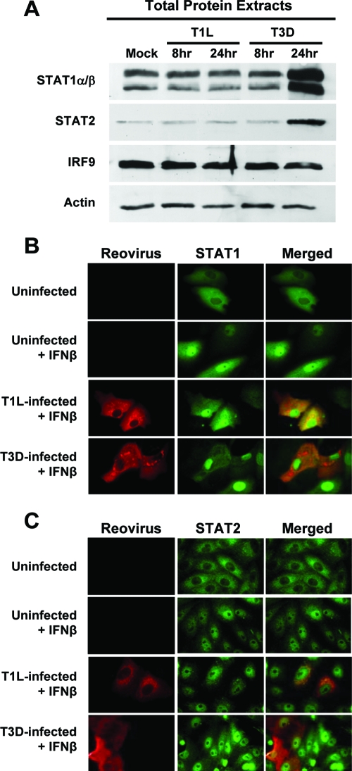
FIG. 5.
T1L does not degrade or inhibit translocation of STATs. (A) L929 cells were infected with either T1L or T3D at an MOI of 25 PFU per cell, and at the indicated times postinfection, total cellular protein was analyzed for STAT1, STAT2, and IRF9 by immunoblotting. Results are representative of two independent experiments. (B and C) Vero cells were infected with either T1L or T3D at an MOI of 25 PFU per cell and incubated for 20 h. Cells were either mock treated or treated with human IFN-β at 1,000 U per ml for 30 min. Cells were fixed and stained with antisera specific for T1L and T3D (B) (red) or monoclonal antibodies specific for reovirus σ1 and σ3 (C) (red) and either STAT1 (B) (green) or STAT2 (C) (green). A yellow color in the merged images indicates overlap of red and green pixels. Representative fields of view show infected and uninfected cells in the same field. STAT nuclear translocation was observed in every T1L- and T3D-infected cell.

