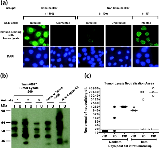
FIG. 3.
Anti-Ad5 Ab are present inside tumors. Tumors were isolated from immunized and nonimmunized hamsters from the experiments in Fig. 2 at 7 and 13 days after the first i.t. injection of vector. The tumors were homogenized in PBS, and the supernatants were used for the assays. (a) Immunofluorescence. INGN 007-infected (20 PFU/cell for 24 h) or uninfected A549 cells were used for an immunofluorescence assay with tumor lysates (isolated 7 days following the first injection of INGN 007). Alexa Fluor 488-conjugated goat anti-hamster IgG was used to detect the hamster Ab against the vector. (b) Western blotting. INGN 007-infected (I) or uninfected (U) A549 cell lysates (multiplicity of infection of 50 PFU/cell) were electrophoresed and transferred to a membrane. The membrane was cut into strips, and a Western blot was performed with 1:500 diluted tumor lysates (isolated 7 days after the first INGN 007 injection) from three different animals in the Imm+007 group, a serum from the immunized group, and a rabbit anti-capsid Ab as indicated. (c) Ad neutralization assay. The tumor lysate was assayed for the presence of anti-Ad NAb.

