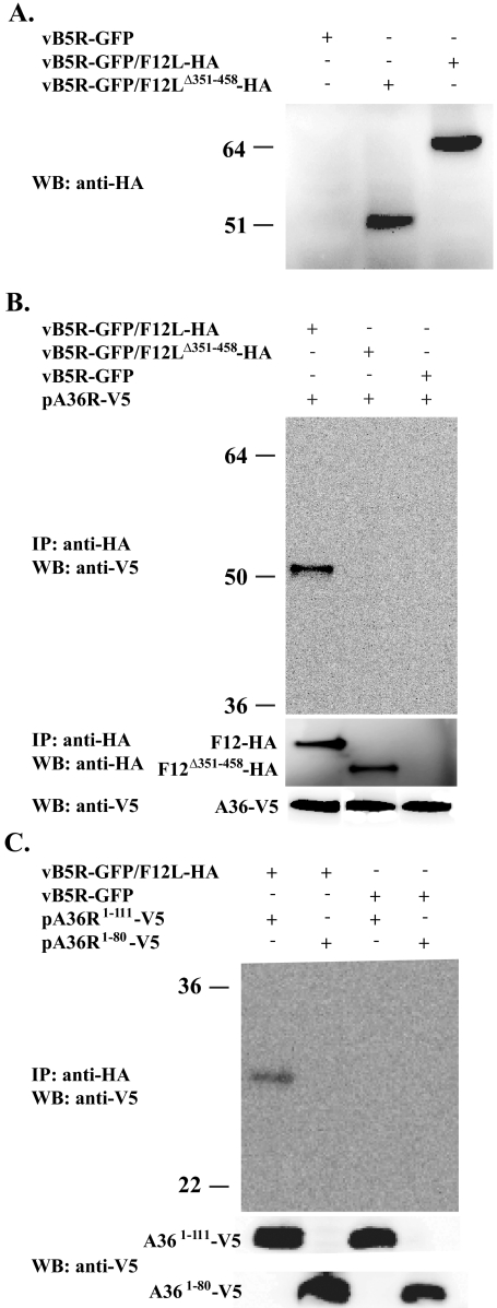
FIG. 3.
Coimmunoprecipitation. (A) Lysates from cells infected with the indicated viruses were analyzed by Western blotting using an anti-HA MAb. HeLa cells were infected with the indicated viruses followed by transfection with the indicated plasmids. Lysates from the infected/transfected cells were immunoprecipitated with anti-HA MAb. Immune complexes were separated by SDS-PAGE, blotted to nitrocellulose, and probed with either an anti-V5 HRP-conjugated MAb (B and C) or an anti-HA MAb (B middle). The bottom panels show the expression of A36-V5 proteins. The positions and masses (in kilodaltons) of markers are indicated on the left.

