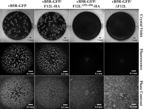
FIG. 4.
Plaque phenotypes. Monolayers of BS-C-1 cells were infected with the indicated recombinant viruses. At 72 h p.i., individual plaques were imaged by fluorescence (middle) and phase-contrast microscopy (bottom). After microscopy, cells were stained with crystal violet, and the entire well was imaged (top).

