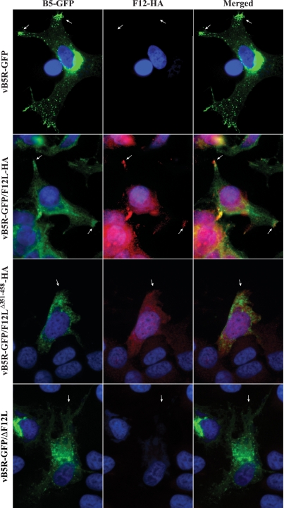
FIG. 6.
Localization of B5-GFP and F12-HA in infected cells. HeLa cells infected with indicated viruses were permeabilized and stained with an anti-HA MAb followed by a Texas Red-conjugated anti-mouse MAb (red). Cells were imaged by fluorescence microscopy. Green represents B5-GFP fluorescence, blue shows DAPI staining, and yellow represents the overlap of green and red fluorescence. Arrows point to the vertices of the cells.

