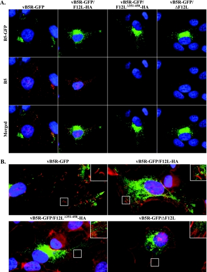
FIG. 7.
(A) Surface staining of B5 on infected cells. HeLa cells were infected with the indicated viruses. At 24 h p.i., unpermeabilized cells were stained with an anti-B5 MAb, followed by a Texas Red-conjugated anti-rat MAb (red), and imaged by fluorescence microscopy. Green represents B5-GFP fluorescence, blue is DAPI staining, and yellow represents overlap of green and red fluorescence. (B) HeLa cells infected with indicated viruses were permeabilized, stained with Alexa Fluor 633 phalloidin (red), and imaged by fluorescence microscopy. Green represents B5-GFP fluorescence, and blue is DAPI staining. Boxed regions are enlarged to show structural details.

