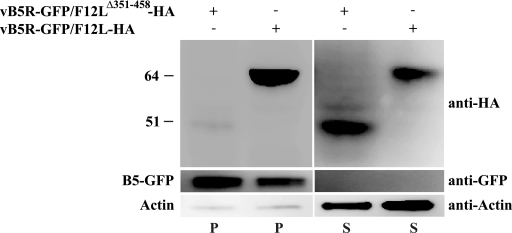
FIG. 9.
Analysis of membranes for the presence of F12. Cells infected with the indicated viruses were harvested 24 h p.i. and lysed. Lysates were centrifuged to pellet membranes, and the resulting pellets (P) and supernatants (S) were analyzed by Western blotting for the presence of F12-HA, B5-GFP, and actin with the indicated MAbs. The positions and masses (in kilodaltons) of markers are indicated on the left.

