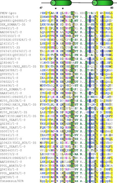
FIG. 1.
Alignment of FMDV Lpro partial amino acid sequence. Lpro protein sequence was aligned to all available sequences utilizing SMART software. Depicted are sequences in single letters between amino acids 47 and 88, and a schematic diagram displays the approximate location of predicted α-helices (green ovals). Asterisks mark the location of amino acids targeted by mutagenesis. Summary of color coding including consensus sequence: + (positive sign in blue), positive charged amino acids (H, K, and R); b (lowercase letter “b” highlighted in yellow), amino acids with a large or bulky side chain (E, F, H, I, K, L, M, Q, R, W, and Y); c (lowercase letter “c” in pink font), charged amino acids (D, E, H, K, and R); l (lowercase letter “l” highlighted in yellow), aliphatic amino acids (I, L, and V); p (lowercase letter “p” in blue font), polar amino acids (C, D, E, H, K, N, Q, R, S, and T); s (lowercase letter “s” in green font), amino acids with a small side chain (A, C, D, G, N, P, S, T, and V); and gray highlighted, amino acids with ≥60% of the homology in the alignment.

