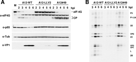
FIG. 7.
Processing of cellular proteins during infection with WT and mutant FMDV. (A) LF-BK cells were infected with WT (A12-WT), leaderless (A12-LLV2), and SAP mutant (A12#49) at MOIs of 10 for 6 h. At the indicated times, cytoplasmic extracts were prepared and analyzed by Western blotting using rabbit polyclonal Ab anti-eIF-4G (p220), rabbit polyclonal Ab anti-p65/RelA (RB-1638), mouse MAb 6HC4 (VP1), and mouse MAb anti-tubulin-α (Ab-2 MS-581). CP, p220 cleavage products. (B) Radioimmunoprecipitation of FMDV-infected cell lysates at different times postinfection. [35S]methionine-labeled cell lysates from FMDV-infected LF-BK cells were immunoprecipitated with serum from a convalescent bovine. Samples were resolved by sodium dodecyl sulfate-polyacrylamide gel electrophoresis and developed by autoradiography.

