FIG. 1.
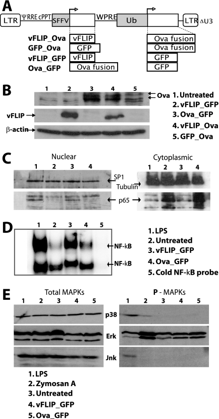
Lentiviral vectors, transgene expression, and cell signaling. (A) Dual-promoter lentivectors expressing vFLIP and an ovalbumin antigen (Ova, ovalbumin epitopes fused to the MHC class II invariant chain) with control vectors. LTR, long terminal repeat; ψ, packaging signal; RRE, Rev-responsive element; cPPT, central polypurine tract; SFFV, spleen focus-forming virus promoter; WPRE, woodchuck hepatitis virus posttranscriptional regulatory element; Ub, human ubiquitin C promoter. (B) Immunoblot showing Ova and vFLIP expression 48 h after transduction of 293T cells at an MOI of 10. Twenty micrograms of protein was loaded per lane. Results from one representative experiment of three are shown. (C) BM-derived DCs were harvested 4 days after transduction. Immunoblots of nuclear samples (13 μg/lane) and cytoplasmic samples (26 μg/lane) were probed with Abs for tubulin, SP1, and p65. Tubulin was not detected in the nucleus, and SP1 was not detected in the cytoplasm (not shown). A repeat experiment showed similar results. (D) Electrophoretic mobility shift assay analysis with DC nuclear extracts made at 5 days; NF-κB binding activity in all DC groups was competed with cold probe (lane 5). Results from one representative experiment of three are shown. (E) Immunoblots for phosphorylated and total MAPKs using DC cell extracts prepared 5 days posttransduction. Samples (15 μg/lane) were probed for P-p38, P-ERK, and P-JNK and for total p38, ERK, and JNK. The experiment shown was repeated two or three times for each MAPK, confirming these results.

