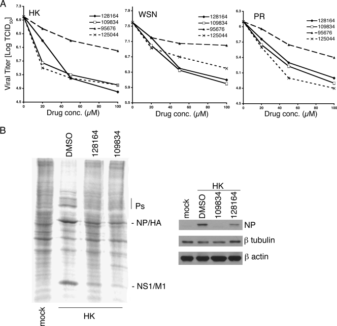
FIG. 3.
Inhibition of virus replication in MDCK cells. (A) Cells were infected with the indicated viruses at an MOI of 0.1 and treated with drug or 1% DMSO (shown as 0 μM) starting 1 h postinfection. The DMSO concentration for all drug-treated cultures was 1%. After incubation for 48 h, the supernatants were collected and analyzed for TCID50 by the method of Reed and Muench (57). (B) Protein expression in infected cells. In the left panel, MDCK cells were infected with A/HK/19/68 at an MOI of 0.1 for 24 h, in the presence or absence of the indicated compounds at 50 μM. The cells were labeled with [35S]methionine and [35S]cysteine for the final hour of infection. Cell lysates were analyzed by SDS-PAGE and visualized by using a phosphorimager. In the right panel, infected cells were analyzed for total NP protein expression by Western blotting. Blots were also probed for β-tubulin and β-actin as loading controls.

