
FIG. 2.
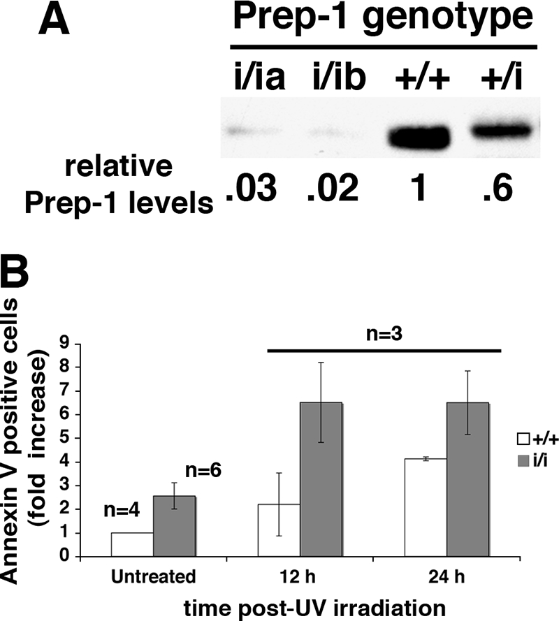
MEFs from Prep1i/i embryos are more apoptotic and more sensitive to UV irradiation than MEFs from WT embryos. (A) SDS-8% PAGE immunoblots of nuclear extracts of MEFs from E14.5 Prep1+/+ (+/+), Prep1+/i (+/i), and Prep1i/i (i/ia and i/ib) littermate embryos. Actin-normalized densitometric quantitation is shown under each lane. The Prep1 content of Prep1+/+ MEFs was arbitrarily set to 1. (B) Prep1+/+ and Prep1i/i MEFs were irradiated with UV light (UV-C; 254 nm; 1,000 J/m2). The number of early apoptotic cells was determined by flow cytometry using annexin V binding. WT untreated cells were given an arbitrary value of 1. n, number of embryos whose MEFs were analyzed. The error bars indicate standard deviations.