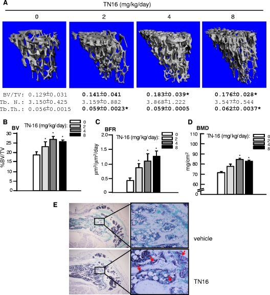
FIG. 3.
Effects of inhibition of microtubule assembly on trabecular bone formation. Three-month-old ICR Swiss mice were injected intraperitoneally with TN16 at doses of 0, 2, 4, and 8 mg/kg/day (50 μl per injection twice a day for 2 days) and sacrificed 1 month later. The mice were also injected with tetracycline and calcein at 10 days and 6 days before sacrifice, respectively. At sacrifice, the tibiae were harvested for analyses. (A) MicroCT analysis. Tibial metaphysis was scanned by the ScanCO μCT 40 scanner with 417 slices. Two hundred slices just beneath the growth plate were analyzed using the ScanCO 40 software program to measure BV/TV, Tb.N., Tb.Th., and Tb.Sp. and to obtain high-resolution images of tibial trabecular bones. (B, C, and D) Tibial histomorphometry. Paraffin-embedded tibiae from TN16-injected mice given the above doses were processed and stained with H&E. BV/TV was measured on the trabecular area of these sections (B). BRF of tibia trabecular bones were measured by using double fluorescence of tetracycline and calcein (C). BMD of tibial metaphysis was measured using a PIXImus bone densitometer (D). (E) BMP-2 in situ hybridization. Hybridization was performed on sections of the femorae from the same mice using a mouse BMP-2 probe. Five to eight mice per group were used. *, P < 0.05 versus results for control.

