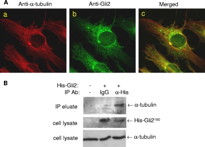
FIG. 4.
Interaction between microtubules and Gli2. (A) Immunofluorescence colocalization of microtubules and Gli2. 2T3 cells cultured in eight-well chamber slides were fixed and incubated with a 1:50 dilution of mouse anti-α-tubulin (Aa) or rabbit anti-mouse Gli2 (Ab) or both antibodies (Ac) and then with the appropriate secondary antibodies against rabbit and mouse IgG, conjugated with FITC and Cy3, respectively. Fluorescence on the mounted slides was captured with a confocal microscope system. (B) Immunoprecipitation of microtubules and Gli2.Cell lysates from 2T3 cells transfected with a His-Gli2 vector were incubated with rabbit Omni-probe anti-His antibody (lane 3), IgG (lane 2), or no Ab (lane 1) and then loaded onto an immobilized protein G column. The eluted immunoprecipitated complex was separated by SDS-PAGE and blotted with anti-α-tubulin antibody (upper panel). The expression of His-Gli2 (middle) or α-tubulin (bottom) as a control in cell lysates was detected by blotting with anti-His or anti-α-tubulin antibody (middle).

