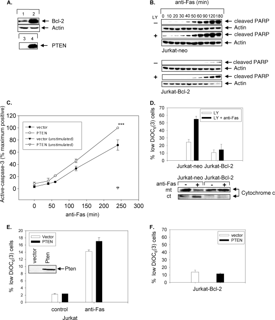
FIG. 3.
PTEN expression or PI3K inhibition sensitizes PTEN-deficient type II Jurkat cells to mitochondrially independent Fas-induced apoptosis. (A) Bcl-2 protein expression in Jurkat-neo (lane 1) and Jurkat-Bcl-2 (lane 2) cells. PTEN protein expression in Jurkat-Bcl-2 vector-transfected cells (lane 3) and PTEN-transfected Jurkat-Bcl-2 cells (lane 4) is also shown. (B) Time course of Fas-induced apoptosis in Jurkat-neo (upper panels) or Jurkat-Bcl-2 (lower panels) cells pretreated with 10 μM LY294002 (+) or DMSO (−) for 2 h. A Western blot of cleaved PARP is shown. (C) Percentage of cells expressing intracellular active caspase-3 in Jurkat-Bcl-2 cells transiently transfected with either vector alone or PTEN following stimulation with anti-Fas for various lengths of time, as indicated, or after being left unstimulated for 4 h. Data represent the means and SEM for cells expressing active caspase-3, presented as the percentages of maximum achievable apoptosis in experiments performed in triplicate. ***, P < 0.0005. (D) Changes in ΔΨm and levels of mitochondrial (mt) and cytoplasmic (ct) cytochrome c from Jurkat-neo and Jurkat-Bcl-2 cells pretreated with LY294002 (10 μM) prior to stimulation in the absence or presence of anti-Fas (CH11) antibody (250 ng/ml) for 90 min. (E) Effect of transient expression of PTEN on ΔΨm of Jurkat cells. A PTEN immunoblot is shown (inset). (F) Absence of ΔΨm in Jurkat-Bcl-2 cells transiently transfected with vector or PTEN followed by treatment with anti-Fas (CH11) for 4 h. Cells were analyzed by flow cytometry for ΔΨm, using the fluorochrome DiOC6 (3). The mean (%) and SEM for cells with low ΔΨm are shown. Experiments were performed in triplicate.

