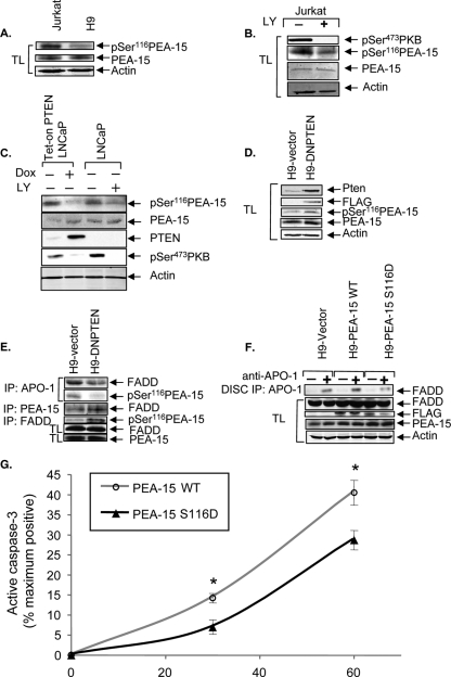
FIG. 5.
PTEN regulates phosphorylation of PEA-15, and overexpression of a phosphomimetic mutant of PEA-15 in type I H9 cells causes them to undergo type II-like kinetics of caspase-3 activation. (A) PEA-15 expression and phosphorylation status in type II Jurkat and type I H9 cells. The levels of endogenous phospho-[Ser116]-PEA-15 and total PEA-15 in whole-cell lysates from cells serum starved for 4 h are shown. The blot was stripped and reprobed for actin as a loading control. (B) The effects of PI3K inhibition on the phosphorylation status of PKB and PEA-15 were monitored by immunoblotting for phospho-[Ser473]-PKB and phospho-[Ser116]-PEA-15 in whole-cell lysates (TL) of Jurkat cells treated with DMSO or LY294002 (40 μM) for 4 h. The blot was stripped and reprobed to demonstrate the level of total PEA-15. The same blot was probed with anti-actin as a loading control. (C) Tet-on PTEN LNCaP cells were treated with or without Dox (4 μg/ml) for 48 h in medium containing 10% serum. Serum was then withdrawn for an additional 48 h. Parental LNCaP cells were treated in parallel with either DMSO or LY294002 (40 μM) for 48 h in the absence of serum. The levels of PTEN, phospho-[Ser473]-PKB, phospho-[Ser116]-PEA-15, and total PEA-15 in whole-cell lysates are shown. Actin was used as a loading control. (D) DN PTEN expression in H9 cells. Lentivirus-driven DN PTEN expression was detected by immunoblotting with anti-PTEN and anti-FLAG antibodies. The levels of total PEA-15 and phospho-[Ser116]-PEA-15 in H9-DN PTEN cells compared to those in vector controls are shown. Actin was used as a loading control. (E) DISC-associated FADD in H9-vector and H9-DN PTEN cells. FADD-associated PEA-15 and total FADD in H9-DN PTEN cells are shown compared to those in H9-vector cells. Phospho-[Ser116]-PEA-15 associated with FADD and total PEA-15 levels in H9-vector and H9-DN PTEN cells are also shown. (F) DISC-associated FADD in H9-vector, H9-PEA-15 WT, and H9-PEA-15 S116D cells. The levels of exogenous PEA-15 WT and PEA-15 S116D were demonstrated by reprobing the blot with anti-FLAG. The levels of total PEA-15, FADD, and actin are shown to demonstrate equal loading of whole-cell lysates. (G) Time course (0 to 60 min) of apoptosis induced by anti-Fas (0.5 μg/ml), as measured by flow cytometric analyses of intracellular active caspase-3 in H9 cells stably expressing PEA-15 WT or PEA-15 S116D. Data represent the means and SEM for cells expressing active caspase-3, presented as percentages of the maximum achievable apoptosis in experiments performed in triplicate and corrected by subtracting the level of spontaneous apoptosis of untreated control cells. *, P < 0.05.

