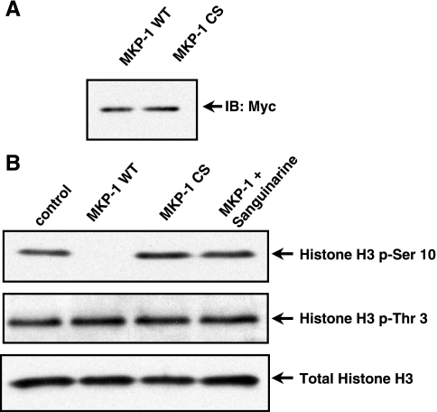
Fig. 4.
MKP-1 dephosphorylates histone H3 at the Ser-10 position in vitro. A: immunoblot analysis of purified MKP-1 (WT) and (CS). B: phosphorylated histones (5 μg) were isolated by acid extraction then acetone precipitated. Histones were subsequently incubated with immunoprecipitated MKP-1 (WT) or (CS) for an in vitro dephosphorylation assay. Histone H3 (Ser-10) and histone H3 (Thr-3) phosphorylation were analyzed by immuno-blot. MKP-1 (WT) dephosphorylated the histone H3 (Ser-10), but not histone H3 (Thr-3). Sanguinarine (SA) blocked MKP-1 dephosphorylation of histone H3 (Ser-10) in vitro. Total histone H3 was used as a loading control.

