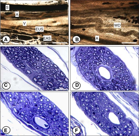
Fig. 4.
Pudendal nerve in the ischiorectal fossa 3 mm distal from the crush site 10 days after injury (A and B) and the motor branch of the pudendal nerve 3 mm distal to the nerve crush site 21 days after injury [control (C), VD (D), PNC (E), and PNC+VD (F)]. A, artery; S, sensory branch; V, vein; EAS, external anal sphincter branch; EUS, EUS branch; WD, Wallerian degeneration identified by clumps of osmophilic ovoids. Stain: lightly osmicated (A and B), toluidine blue (C–F). Bar = 1.5 mm (A), 0.5 mm (B), 10 μm (C–F).

