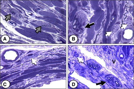
Fig. 5.
Nerve fascicles near the EUS 21 days after injury. A: control. B: VD. C: PNC. D: PNC+VD. Gray arrows, normal-appearing fascicles; white arrows, nonmyelinated axons; black arrows, degenerated fascicles, defined as having waviness of axons, evidence of myelin breakdown debris, and/or increased endoneurial cellularity. V, cytoplasmic vacuoles. Stain: toluidine blue. Bar = 10 μm.

