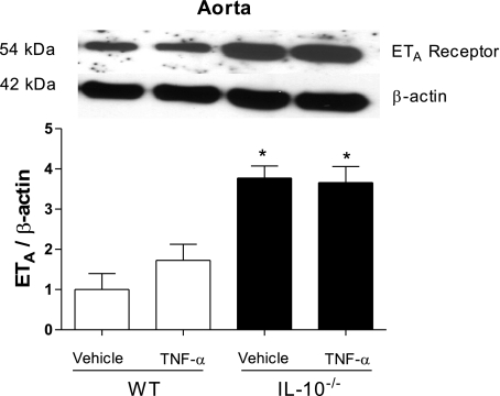
Fig. 4.
ETA receptor expression is augmented in vessels from IL-10−/− mice. Top: representative immunoblots for ETA receptor and β-actin expression in murine aorta. Bottom: corresponding bar graphs demonstrating regulatory role of IL-10 in ETA receptor expression. Densitometric analysis was performed in samples from vehicle- and TNF-α-infused WT and vehicle- and TNF-α-infused IL-10−/− mice. Values, expressed in arbitrary units, are means ± SE (n = 5 experiments) and were normalized to β-actin protein expression. *P < 0.05 vs. respective control (i.e., WT).

