Fig. 7.
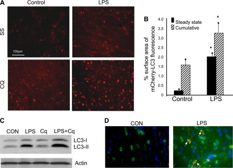
LPS stimulated autophagic flux and upregulated cathepsin D in vivo. Hearts of mCherry-LC3 transgenic mice were harvested 4 h after LPS injection (1.5 mg/kg ip injection) with or without concurrent administration of chloroquine (CQ) to prevent lysosomal degradation of autophagosomes. A: fluorescence micrographs of heart tissues under indicated conditions. Bright dots indicate autophagosomes. B: quantitative analysis of mCherry puncta from each condition. *P < 0.001 compared with control. C: immunoblot of LC3 in heart tissue lysates prepared from mice treated as described above. D: immunodetection of cathepsin D in sections of mCherry-LC3 transgenic mouse hearts treated as above (no CQ). Cathepsin D (green) colocalized with mCherry-LC3 puncta (red) as indicated by arrows. SS, steady state.

