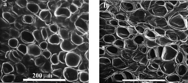
Figure 1.
(a) A micrograph, taken in an environmental scanning electron microscope under conditions of saturated vapor pressure, of the cellular structure of a fresh carrot. (For further details, see Experimental section). The structure has a relatively low volume fraction of solid, in the manner of a classical foam. The concentration of material into edges rather than faces is not very pronounced. (b) The same region under 60% nominal compression. Cells are not distorted uniformly, as the weakest ones burst first and collapse. The partial pressure of water vapor in the specimen chamber was lowered slightly to drive off the excess fluid released from bursting cells, as this would obscure the image.

