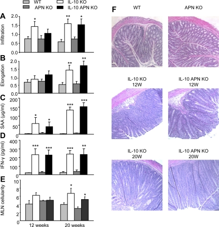
Fig. 1.
Inflammatory infiltrate, colonic hyperplasia, and serum markers of inflammation. Inflammatory cell infiltration (A) and crypt elongation (B) were evaluated in colonic tissue at week 12 and 20. Levels of serum amyloid A (SAA; C) and IFN-γ (D) were measured in serum at the same time points. E: cellularity of the mesenteric lymph nodes. F: representative histological samples of colonic tissue from the indicated groups. Data are means ± SE. N = 10 for wild-type (WT) and IL-10 knockout (KO) mice and N = 14 for adiponectin (APN) KO and double IL-10 APN KO mice. *P < 0.05, **P < 0.01, and ***P < 0.001 WT vs. IL-10 KO mice and APN KO vs. double IL-10 APN KO mice.

