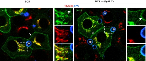
Fig. 3.
Trafficking of N41S, a disease-causing mutation in ATP7B, borders on being completely defective. Polarized WIF-B cells were infected with GFP-N41S-ATP7B, cultured overnight in 10 μM BCS, then kept in BCS or incubated in 10 μM Cu(II) for 4 h, fixed, stained with antibodies to the indicated organelle markers, and imaged by confocal microscopy. Arrows, basolateral membrane; arrowheads, apical membrane; asterisks, apical lumen. Boxed regions are enlarged at the right of each panel.

