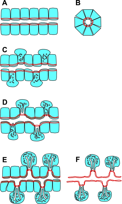
Fig. 10.
Cartoon depicting an hypothesis for morphogenesis of gastric glands. All the cells in gastric glands are derived from stem cells that reside in the isthmus of the gland. The differential patterns for the staining of ZO-1 and moesin in the neck and the body of the glands implicate an interesting course for the continued development of glandular morphology in both space (isthmus to base) and time (shown here as the progressive change from A to D). A and B show respective longitudinal and cross-sectional regions from the isthmus where all cells are undifferentiated. Cytoplasmic matrix of cells is shown in light blue; red lines represent tight junctions with the double red line taking artistic license to indicate staining around the entire periphery of each cell. In early development (C), preparietal cells differentiate from stem cells initiating organellar volume expansion largely projected toward the glandular periphery. With further differentiation (D), parietal cells migrate toward the base of the gland, continuing to grow and expand in the peripheral direction and forming intracellular canaliculi to expand their apical membrane surfaces. This retreat of parietal cells from the lumen of the glands is likely to exert a pulling effect on the gland lumen, making secondary indentations to further expand total glandular luminal secretory surface area (E). The replication, basal migration, and the growth of mucous neck cells and then differentiation into chief cells, in concert with parietal cell development, facilitate the formation of the mature glandular morphology. In F all cells other than parietal cells have been deleted to emphasize the gland luminal structure including the surface indentations projecting toward parietal cells.

