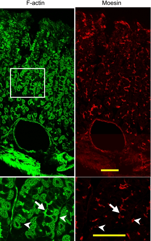
Fig. 8.
Moesin is detected on both endothelial cells and the luminal surface of cells in the base of gastric glands. Cryosections 15 μm thick were prepared from fresh stomach tissue for immunofluorescence staining with mouse anti-moesin antibody followed by the Alexa Fluor 555-conjugated donkey anti-mouse antibody and FITC-conjugated phalloidin. Bottom: higher magnification of the boxed area in the top. Note the moesin staining of a large blood vessel (close to the muscularis externa) in the top. Arrows point to the lumen of the gastric gland (note the moesin staining of the gland lumen) whereas arrowheads point to moesin staining in the endothelial cells. Bar = 50 μm in all cases.

